STED Microscopy with Supercontinuum Lasers

STED microscopy enables high-resolution imaging beyond the diffraction limit using NKT Photonics' SuperK and KATANA HP lasers for flexible pulsed excitation and synchronized depletion.

STED Microscopy

STED microscopy is widely used to study luminescent samples with a high spatial resolution far below the diffraction barrier in the fields of biology, medicine as well as materials science. Therefore, in a confocal laser scanning microscope, the sample is excited with a diffraction-limited, pulsed laser, followed by a doughnut-shaped second laser pulse which is red-shifted with regard to the emission spectrum of the chromophore.

This document provides a quick overview of the STED technique introduced by Stefan W. Hell in 1994. NKT Photonics’ SuperK and KATANA HP lasers are a perfect combination to realize flexible pulsed excitation as well as synchronized depletion within the visible and near-infrared range.
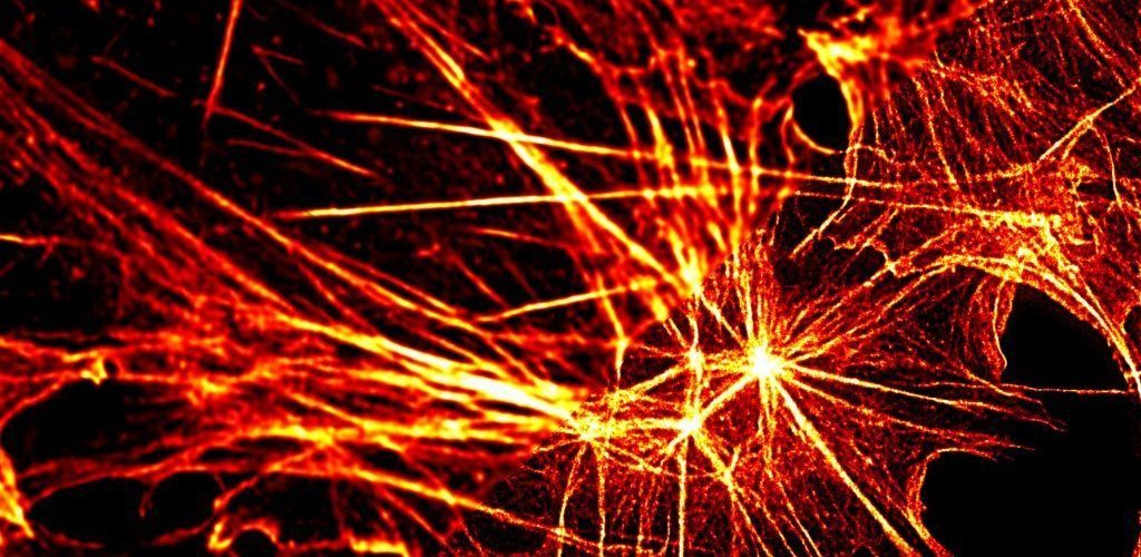
STED illumination

STED microscopy excitation with SuperK laser and SuperK SELECT
STED microscopy excitation using SuperK laser and SuperK SELECT, demonstrating high-resolution imaging capabilities.
STED microscopy depletion with SuperK laser and SuperK SELECT
STED microscopy depletion using SuperK laser and SuperK SELECT, demonstrating high-resolution imaging capabilities.
This image illustrates STED microscopy using the SuperK laser and SuperK SELECT. It highlights the advanced capabilities of NKT Photonics' lasers in providing high-resolution imaging for biological and medical applications. The SuperK laser, combined with the SuperK SELECT, offers tunable laser sources ideal for precise excitation and depletion in fluorescence microscopy, enabling superior imaging beyond the diffraction limit.
STED microscopy using SuperK laser and SuperK SELECT, demonstrating high-resolution imaging capabilities.

This leads to a depletion of the outer ring of the confocal excitation volume.  The remaining fluorescence after the depletion pulse is therefore only emitted from a shrunk region in the center of the excitation volume.

Pulse scheme for STED microscopy using SuperK laser and SuperK SELECT
Pulse scheme for STED microscopy using SuperK laser and SuperK SELECT, demonstrating high-resolution imaging capabilities.

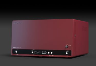
The SuperK Supercontinuum lasers deliver a continuous spectrum over the visible (Vis) and near-infrared (nIR) range, with excellent single-mode beam profile (M2 < 1.1) and picosecond (ps) pulse duration. In combination with our filter technology, it can be transformed into a tunable laser source, allowing optimized excitation of every chromophore absorbing in the Vis and nIR regions.
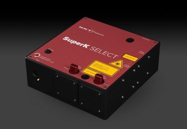
Multiline filter transmission of supercontinuum light from SuperK laser through SuperK SELECT for STED microscopy
Multiline filter transmission of supercontinuum light from SuperK laser through SuperK SELECT, showcasing high-resolution imaging capabilities for STED microscopy.

The Pulsed Diode Lasers are available at various wavelengths within the Vis-nIR and offer high pulse energy at ps pulse duration making them ideal for STED depletion.

Spectral power density of SuperK Extreme EXW-12 and available wavelengths for KATANA HP lasers
The spectral power density of the SuperK Extreme EXW-12 (blue), available wavelength for KATANA HP lasers (red).

In addition to using the proper combination of wavelengths and laser power levels, in STED microscopy it is also crucial to precisely adjust the excitation and depletion laser pulses, to synchronize for efficient depletion of the excited chromophores at the beginning of each fluorescence cycle.

NKT Photonics’ mode-locked Supercontinuum lasers are equipped with a NIM trigger output and built-in adjustable trigger delay as a standard feature. The KATANA HP lasers can run as a slave on external trigger input, which allows for simple software-controlled adjustment of the excitation and depletion pulses.
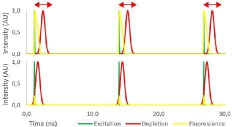
STED microscopy with SuperK CHROMATUNE tunable laser for high-resolution imaging
Explore the capabilities of STED microscopy using the SuperK CHROMATUNE tunable laser for precise and high-resolution imaging.

In the experiment, the resolution is mainly limited by the photophysics of the chromophore used. That is why new fluorescent labels are developed rapidly. Consequently, the flexibility of a Supercontinuum laser to freely choose the excitation wavelength helps to prepare for upcoming labels in the VIS-nIR.

The combination of the SuperK platform with the KATANA HP laser family becomes a turnkey solution for the most flexible and modular implementation of STED microscopy on the market successfully proven through multiple installations worldwide.

Download this article as pdf

Suitable hardware configuration for STED microscopy:

Supercontinuum excitation:

Pulsed depletion:

Optical Coherence Tomography (OCT) enables cross-sectional or 3D imaging of subjects under investigation.

This provides a significant advantage over alternative microscopy techniques, which are typically limited to examining surface or shallow layers.

Cross-sectional and 3D imaging are vital for a wide range of applications, from analyzing tissue in medical contexts to visualizing sub-micron structures in manufacturing.

The principle of OCT imaging was first demonstrated in 1991 by Professor Huang et al. A comprehensive overview of its principles and applications has been provided by Professor Drexler from the Medical University of Vienna and Professor Fujimoto from MIT in Optical Coherence Tomography: Technology and Applications.

Over the past 30 years, OCT has become an indispensable imaging tool in ophthalmology, particularly for the detailed analysis of the retina and surrounding tissues.

However, OCT applications are not confined to ophthalmology. A growing body of research focuses on exploring OCT in diverse fields beyond eye care.

SuperK supercontinuum lasers offer several key parameters critical to Ultra-High-Resolution OCT (UHR-OCT):

  • Extremely broad optical bandwidths
  • Excellent spatial coherence
  • High optical power density

The use of SuperK sources in OCT applications is expanding rapidly, reflecting their growing importance in advancing this transformative imaging technology.

OCT in a nutshell

OCT relies on interferometry. Light from one arm is reflected or scattered by the subject under investigation and interferes with light from a reference arm.

Both light beams originate from the same source. Interference occurs when the difference in path lengths between the two arms is within the coherence length of the optical signal.

This coherence gating allows the detection system to distinguish reflections from closely spaced reflectors, enabling high-resolution imaging.

The sensitivity of OCT is exceptionally high, allowing it to detect even weak signals from sub-surface reflections. This capability enables cross-sectional imaging, similar to ultrasound, but with significantly higher resolution. Imaging depths of several millimeters into tissue can be achieved.

Practical realizations of OCT

There are several practical implementations of OCT, each with unique characteristics:

  • Time-domain OCT (TD-OCT)
    The reference mirror moves, enabling coherence gating at different depths in the sample arm. This was the first implementation of OCT and remains relevant in certain applications, such as Full-Field OCT, where the interference pattern for a full 2D array is simultaneously detected by a 2D detector array (e.g., CCD or CMOS).
  • Spectral-domain OCT (SD-OCT)
    Also known as Fourier-Domain OCT (FD-OCT), this approach uses a fixed reference mirror. The interference pattern is detected spectrally and converted to spatial information via Fourier transformation.
  • Spectrometer-based OCT (Sp-OCT)
    A broadband source, such as a SuperK source, generates the interference spectrum, which is detected by a high-speed spectrometer. This typically involves several thousand pixels and sub-nanometer optical resolution.
  • Swept-source OCT (SS-OCT)
    A tunable light source rapidly scans the relevant spectral range, and the spectral response of the interferometer is detected using a single or balanced detector.

Each of these techniques offers distinct advantages and disadvantages, making them more or less suitable for specific applications.

SuperK supercontinuum white light lasers are versatile and can be used across all these OCT implementations. For SS-OCT, a SuperK source can be combined with a rapidly scanning bandpass filter to effectively sweep the center wavelength. However, most SuperK sources have been applied to SD-OCT based on spectrometer detection (Sp-OCT).

Low noise ensures high-contrast images

The SuperK FIANIUM supercontinuum fiber laser series has the lowest noise on the market. Optimized for low-noise performance, it gives high-contrast low-noise images in OCT systems. Its performance matches that of bulky Ti:Sapphire lasers.

Below, you can see two OCT images of a human eye. The image on the right is recorded using a SuperK EXTREME OCT source (now replaced by the SuperK FIANIUM OCT), while the image on the left is obtained using a Ti:Sapphire laser. Both images are recorded by Angelika Unterhuber from Prof. Dr. Wolfgang Drexlers group at Medical University Vienna.

SuperK white light lasers for OCT

Explore how others have used SuperK supercontinuum white light lasers for OCT in these white papers.


Articles and background info

Get some background information.