Techniques used in ophthalmic imaging
Optical Coherence Tomography (OCT)
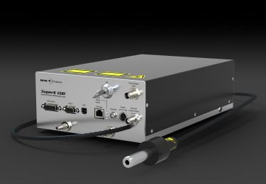
Learn more about the basic principles of supercontinuum powered OCT.
| Publications | Summary |
| Ji Yi, 2013; Visible-light optical coherence tomography for retinal oximetry | Using SuperK as OCT light to extract hemoglobin oxygen saturation (sO2) in individual retinal vessels. They established a comprehensive analytical model to describe optical absorption, optical scattering, and blood cell packing factor in the whole blood and fit the acquired vis-OCT signals from the bottom of each imaged vessel. |
| Ji Yi, 2012,; Structured interference optical coherence tomography | SuperK was used for illumination in order to get an axial resolution of ~1.5μm in air (650-800 nm) |
Scanning Laser Ophthalmology (SLO)
| Publications | Summary |
| Wolf M. Harmening, 2014; Mapping the Perceptual Grain of the Human Retina | Using AOSLO imaging to map the cone mosaic with subcellular resolution. The SuperK was the light source; 842 nm and 543 nm at the same time |
| Francesco LaRocca, 2014; True color scanning laser ophthalmoscopy and optical coherence tomography handheld probe | The SuperK was used as SLO source (430-700 nm) for three colors. True color SLO retina may provide better sensitivity, this higher sensitivity to color variations in the retina, the “true color” SLO could potentially provide earlier detection of age-related macular degeneration (AMD) by earlier visualization of druses, which appears as yellow-white spots in the retina. |
| Drew Scoles, 2013; In vivo dark-field imaging of the retinal pigment epithelium cell mosaic | SuperK was used as the light source, centered either at 565 or 680 nm, with a 10 nm bandwidth |
| Wolf M. Harmening, 2012; Measurement and correction of transverse chromatic offsets for multi-wavelength retinal microscopy in the living eye | SuperK is provided light for the 842 nm imaging channel as well as the red (711 ± 12 nm) and green (543 ± 11 nm) channels used for stimulation |
Articles and background info
- Paper by Adam Wax, Medical Physics Program, Duke University: Assessing hemoglobin concentration using spectroscopic optical coherence tomography for feasibility of tissue diagnostics
- Research article by Adam Wax, Biomedical Engineering, Duke University: Deep tissue imaging using spectroscopic analysis of multiply scattered light
- Paper by Adam Wax: Fourier domain multispectral multiple scattering low coherence interferometry