Lumpectomy is the surgical procedure of choice for the vast majority of breast cancer patients. It is usually conducted in an outpatient setting and carries a low risk of complications. It offers the possibility of complete recovery, and – if we can make this procedure more effective – we can achieve major economic and emotional benefits.
The challenge we face today is that about 1 in 3 patients needs follow-up surgery due to incomplete cancer removal.
To address this issue, a team at a major medical center in Los Angeles, California, developed a technology platform utilizing NKT Photonics aeroPULSE PS UV laser, which is intended to help surgeons during surgery to avoid incomplete tumor excision.
Initial clinical studies show a nearly 100% success rate when compared to pathology in detecting cancer versus normal breast tissue, thus potentially reducing the need for follow up-surgeries.
Battling against incomplete cancer removal
Breast cancer is one of the most common cancers in women today with one out of eight women being diagnosed with the disease in their lifetime. About 75% of patients undergo a lumpectomy, a breast-conserving tumor-removal surgery. With increasing awareness and advances in early-detection schemes, the number of lumpectomy procedures is growing in double-digits year over year.
Unfortunately, about 1 in 3 patients need follow-up surgery because of incomplete tumor removal. This results not only in a high burden on health care systems but also creates anxiety in patients undergoing repeated procedures. New technologies are needed to help surgeons in achieving near-complete tumor excisions and hence significantly reduce the number of follow-up surgeries.
A research team led by Dr. Pramod Butte, together with surgeons and pathologists at a medical center in Los Angeles, California, has developed a new laser-based platform to address this issue. The technology can be deployed during the lumpectomy procedure and aids the surgeon in deciding if sufficient tissue has been removed.
A spin-off company named BlackLight Surgical was founded with the target of bringing this valuable innovation to market by further developing the technology and obtaining necessary regulatory approvals.
The platform has already been tested in a series of clinical studies at local centers. The result: they found an astonishing near 100% success rate compared to pathology in detecting cancer versus normal breast tissue, thus potentially minimizing the need for follow-up surgeries.
How does it work?
During the operation, the surgeon removes the cancerous tissue from the breast, which is immediately scanned by the BlackLight Surgical imaging system. This scan informs the surgeon in under a minute whether cancerous tissue is present on the surface of the excised tissue.
A negative result means that all the cancer was encased inside the removed tissue, and the excision is complete.
A positive result on the surface of the excised tissue indicates that cancer was left behind in the breast.
Today, this would only be identified in a time-consuming histopathological analysis, typically issued days after the procedure and leading to follow-up surgery for the patient.
With the technology from BlackLight Surgical, cancer on the surface can be detected during surgery, and the surgeon can readily decide to remove additional tissue intra-operatively. This tissue, in turn, can be scanned to confirm all cancer has been removed, thus creating the opportunity for a significant reduction in repeat surgeries.

The technology behind the scenes
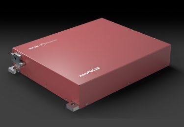
To identify cancerous tissue on the surface of the excised tissue, BlackLight Surgical is utilizing fluorescence lifetime imaging. An ultrafast picosecond pulse from an NKT Photonics aeroPULSE PS UV laser excites specific regions of the specimen.
The emitted autofluorescent traces are analyzed in time and at different wavelengths, see below.

The autofluorescent traces are dependent on the cell type and are different for, e.g., fat cells or stromal cells versus cancer cells. Therefore, careful analysis of the temporal traces at various wavelengths enables the identification of signatures for cancerous cells that are separate from healthy cells.
This principle is paired with a novel imaging set-up for scanning tissue samples during the operation in under a minute.
The output image allows the surgeon to determine, during the operation, whether there is cancer on the surface of the scanned tissue with high accuracy, see photos below (images from bls.life).


The system from BlackLight Surgical uses ultrashort laser pulses from our aeroPULSE PS picosecond UV laser to excite the autofluorescence. Our unique fiber-laser design gives a highly reliable laser performance with a short start-up time and robustness against environmental changes.
These features also allow for the integration into a cart that can be transported between operating rooms. This powerful example shows how our lasers enable technology platforms in clinical applications that will help women affected by breast cancer worldwide.
Future applications
Looking into the future and pending regulatory approvals, this promising technology can also be adapted to other surgeries involving solid tumors such as brain, head & neck, lung, and prostate.