Advanced Imaging with Optical Coherence Tomography
Ultra-high resolution Optical Coherence Tomography (UHR-OCT) is an invaluable tool that enables micron-scale cross-sectional or 3D imaging of a subject. It is relevant for various applications, from analysis of tissue in medical applications to visualization of sub-micron structures in manufacturing.
Optical Coherence Tomography (OCT) enables cross-sectional or 3D imaging of subjects under investigation.
This provides a significant advantage over alternative microscopy techniques, which are typically limited to examining surface or shallow layers.
Cross-sectional and 3D imaging are vital for a wide range of applications, from analyzing tissue in medical contexts to visualizing sub-micron structures in manufacturing.
The principle of OCT imaging was first demonstrated in 1991 by Professor Huang et al. A comprehensive overview of its principles and applications has been provided by Professor Drexler from the Medical University of Vienna and Professor Fujimoto from MIT in “Optical Coherence Tomography: Technology and Applications.“
Over the past 30 years, OCT has become an indispensable imaging tool in ophthalmology, particularly for the detailed analysis of the retina and surrounding tissues.
However, OCT applications are not confined to ophthalmology. A growing body of research focuses on exploring OCT in diverse fields beyond eye care.
SuperK supercontinuum lasers offer several key parameters critical to Ultra-High-Resolution OCT (UHR-OCT):
Extremely broad optical bandwidths
Excellent spatial coherence
High optical power density
The use of SuperK sources in OCT applications is expanding rapidly, reflecting their growing importance in advancing this transformative imaging technology.
OCT in a nutshell
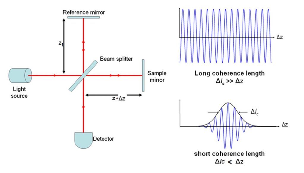
OCT relies on interferometry. Light from one arm is reflected or scattered by the subject under investigation and interferes with light from a reference arm.
Both light beams originate from the same source. Interference occurs when the difference in path lengths between the two arms is within the coherence length of the optical signal.
This coherence gating allows the detection system to distinguish reflections from closely spaced reflectors, enabling high-resolution imaging.
The sensitivity of OCT is exceptionally high, allowing it to detect even weak signals from sub-surface reflections. This capability enables cross-sectional imaging, similar to ultrasound, but with significantly higher resolution. Imaging depths of several millimeters into tissue can be achieved.
Practical realizations of OCT
There are several practical implementations of OCT, each with unique characteristics:
Time-domain OCT (TD-OCT) The reference mirror moves, enabling coherence gating at different depths in the sample arm. This was the first implementation of OCT and remains relevant in certain applications, such as Full-Field OCT, where the interference pattern for a full 2D array is simultaneously detected by a 2D detector array (e.g., CCD or CMOS).
Spectral-domain OCT (SD-OCT) Also known as Fourier-Domain OCT (FD-OCT), this approach uses a fixed reference mirror. The interference pattern is detected spectrally and converted to spatial information via Fourier transformation.
Spectrometer-based OCT (Sp-OCT) A broadband source, such as a SuperK source, generates the interference spectrum, which is detected by a high-speed spectrometer. This typically involves several thousand pixels and sub-nanometer optical resolution.
Swept-source OCT (SS-OCT) A tunable light source rapidly scans the relevant spectral range, and the spectral response of the interferometer is detected using a single or balanced detector.
Each of these techniques offers distinct advantages and disadvantages, making them more or less suitable for specific applications.
SuperK supercontinuum white light lasers are versatile and can be used across all these OCT implementations. For SS-OCT, a SuperK source can be combined with a rapidly scanning bandpass filter to effectively sweep the center wavelength. However, most SuperK sources have been applied to SD-OCT based on spectrometer detection (Sp-OCT).
Low noise ensures high-contrast images
The SuperK FIANIUM supercontinuum fiber laser series has the lowest noise on the market. Optimized for low-noise performance, it gives high-contrast low-noise images in OCT systems. Its performance matches that of bulky Ti:Sapphire lasers.
Below, you can see two OCT images of a human eye. The image on the right is recorded using a SuperK EXTREME OCT source (now replaced by the SuperK FIANIUM OCT), while the image on the left is obtained using a Ti:Sapphire laser. Both images are recorded by Angelika Unterhuber from Prof. Dr. Wolfgang Drexlers group at Medical University Vienna.
SuperK white light lasers for OCT
Explore how others have used SuperK supercontinuum white light lasers for OCT in these white papers.
Shot-noise limited, supercontinuum-based optical coherence tomography by Shreesha Rao D. S., Mikkel Jensen, Lars Grüner-Nielsen, Jesper Toft Olsen, Peter Heiduschka, Björn Kemper, Jürgen Schnekenburger, Martin Glud, Mette Mogensen, Niels Møller Israelsen, and Ole Bang, published in Light: Science & Applications, 10, Article number: 133 , 2021.
Real-time high-resolution mid-infrared optical coherence tomography by Niels M. Israelsen, Christian R. Petersen, Ajanta Barh, Deepak Jain, Mikkel Jensen, Günther Hannesschläger, Peter Tidemand-Lichtenberg, Christian Pedersen, Adrian Podoleanu and Ole Bang published in Nature Light: Science & Applications 8, article 11, 2019.
Recovering distance information in spectral domain interferometry by Adrian Bradu, Niels Møller Israelsen, Michael Maria, Manuel J. Marques, Sylvain Rivet, Thomas Feuchter, Ole Bang, and Adrian Podoleanu, published in Scientific Reports 8, Article 15445, 2018.
Visible-light OCT spectrometer for microvascular oximetry by Gangnus, Sergei V., and Stephen J. Matcher, Proceedings of SPIE, the International Society for Optical Engineering. Society of Photo-Optical Instrumentation Engineers, 2008.