MINFLUX is a relatively new fluorescence microscopy technique that lets you distinguish between molecules just nanometers apart. Until now, MINFLUX is the most precise and most photon-efficient way to localize fluorescent molecules.
In a transatlantic collaboration, two groups of scientists worked together to further improve MINFLUX nanoscopy. They invented p-MINFLUX – the pulsed interleaved MINFLUX.
Super-resolution microscopy for single-molecule spectroscopy and localization
The two groups that invented p-MINFLUX by simplifying the MINFLUX microscope are Professor Philip Tinnefeld’s group at the LMU Munich and Professor Fernando Stefani’s group at the University of Buenos Aires.
Professor Philip Tinnefeld explains: “With p-MINFLUX it will be possible to uncover structures and dynamics at the molecular level that are fundamental for our understanding of energy transfer processes in artificial and natural light-harvesting systems and energy conversion materials as well as in biomolecular reactions.”
To do so p-MINFLUX uses interleaved laser pulses to deliver doughnut-shaped excitation profiles in a precisely defined spatial pattern at high MHz repetition rates.
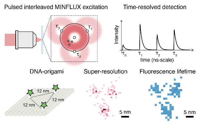
p-MINFLUX lets scientists distinguish different types of molecules and track how each molecule moves independently of other molecules.
To determine the location of a molecule, p-MINFLUX places a laser focus near it. The distance between the molecule and the center of the laser focus can then be deducted from the fluorescence intensity. Triangulation gives the exact position of the molecule. It delivers a 1−2 nm localization precision with only 2000−1000 photon counts.
Furthermore, p-MINFLUX gives access to the fluorescence lifetime information, thus enabling multiplexing super-resolved and lifetime imaging.
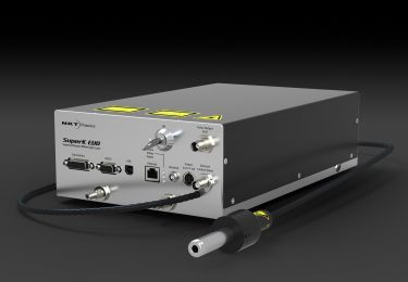
A supercontinuum laser made p-MINFLUX possible
The groups used our high-power, mode-locked SuperK supercontinuum laser system with MHz repetition rate as light sources in their p-MINFLUX microscopes in Munich and Buenos Aires.
Specifications that made the SuperK the ideal laser for p-MINFLUX are:
- Its pulse duration below 100 ps with clean pulse form
- The very low timing jitter
- Its MHz repetition rate
- High pulse energy > 100 pJ
- Wavelength flexibility across the VIS and nIR range
- Good power stability
- Single-mode beam quality with M2 < 1.1
Get all the details in the publication
In their publication in NanoLetters, the scientists give you the background, the principle and experimental set-up, and how to use the p-MINFLUX method.
Meet the authors

Luciano Masullo is a Ph.D. student in Physics at the University of Buenos Aires (Argentina). He studied Physics (B. Sc. and M. Sc.) with a focus on optics and microscopy. His Ph.D. work is being conducted at the Center for Bionanoscience Research (CIBION, Argentina) and focuses on super-resolution microscopy with a molecular-scale resolution, particularly on MINFLUX. He is a strong supporter of open-source science and an enthusiast of Python programming.

Florian Steiner is a postdoc at the Ludwig-Maximilians-Universität München (Germany). He studied physics (B. Sc. and M. Sc.) at the University of Regensburg (Germany) with a focus on solid-state and semiconductor physics. During his Ph.D. studies, he investigated the photophysical properties of conjugated polymers and organic molecules via single-molecule spectroscopy techniques. Since 2018 he has been working in Prof. Philip Tinnefeld’s group at the LMU, focusing on the development and applications of p-MINFLUX.

Philip Tinnefeld has been a Professor of Physical Chemistry at the Ludwig-Maximilians-Universität München (Germany) since 2017. He studied chemistry in Münster and Heidelberg and received his Ph.D. from the University of Heidelberg in 2002. He became an associate professor of biophysics at Ludwig-Maximilians-Universität München after postdoctoral work within research, stays at the UCLA (United States) and Leuven (Belgium), and habilitation in physics at Bielefeld University. In 2010, he was appointed Full Professor of biophysical chemistry at Braunschweig University of Technology. Philip Tinnefeld’s research is inspired by our emerging abilities to study and build matter bottom-up, starting from single molecules. He has contributed to breakthroughs of single-molecule superresolution microscopy and he has combined optical single-molecule detection with DNA nanotechnology for self-assembled, functional devices including energy-transfer switches, calibration nanorulers, nanoadapters, fluorescence signal amplifiers, and molecular force clamps. Dr. Tinnefeld has authored more than 160 publications and patents. He was the initiator of GATTAquant GmbH, the first company commercializing DNA origami applications.

Fernando Stefani is a Full Professor at the Department of Physics at the University of Buenos Aires (Argentina), Deputy Director of CIBION. He studied Materials Engineering at Instituto Sábato – University of San Martín, then obtained his Ph.D. at the University of Mainz (2004), conducting his research at the Max Planck Institute for Polymer Research. He did postdoc work at the Institute for Photonic Science (ICFO) in Barcelona and was then a group leader at the Faculty of Physics of the LMU. In 2010, he was appointed Professor at the University of Buenos Aires. Prof. Stefani’s work focuses on light-matter interactions at the nanoscale with a focus on optical techniques that allow manipulation and visualization of molecules and nanoparticles.